Anatomy of Knee: Understanding the Structure and Function of the Knee Joint

The knee joint is one of the most complex and important joints in the human body. It is responsible for providing stability and mobility to the lower body, allowing us to walk, run, and jump. Understanding the anatomy of the knee joint is essential for proper diagnosis and treatment of knee injuries and conditions. This article will provide an overview of the anatomy of the knee joint, including its structure and function. We will also discuss common knee injuries and conditions, and how they can be treated.

Exploring the Anatomy of the Knee Joint: A Comprehensive Guide

The knee joint is one of the most complex and important joints in the human body. It is responsible for providing stability and mobility to the lower body, allowing us to walk, run, and jump. Understanding the anatomy of the knee joint is essential for proper diagnosis and treatment of knee injuries and conditions. This guide will provide a comprehensive overview of the anatomy of the knee joint, including its structure, ligaments, muscles, and other components.

The knee joint is a synovial joint, which is a type of joint that is surrounded by a capsule filled with synovial fluid. This fluid lubricates the joint and helps to reduce friction between the bones. The knee joint is composed of three bones: the femur, tibia, and patella. The femur is the thigh bone, the tibia is the shin bone, and the patella is the kneecap. These bones are connected by four ligaments: the anterior cruciate ligament (ACL), posterior cruciate ligament (PCL), medial collateral ligament (MCL), and lateral collateral ligament (LCL). The ACL and PCL are located in the center of the knee and provide stability to the joint. The MCL and LCL are located on the sides of the knee and provide stability to the joint when it is bent or twisted.

The knee joint is also supported by several muscles, including the quadriceps, hamstrings, and calf muscles. The quadriceps are located on the front of the thigh and are responsible for extending the knee. The hamstrings are located on the back of the thigh and are responsible for flexing the knee. The calf muscles are located on the back of the lower leg and are responsible for plantar flexing the ankle.

The knee joint is also surrounded by several bursae, which are small sacs filled with fluid that cushion and protect the joint. The most important bursae are the prepatellar bursa, which is located in front of the kneecap, and the infrapatellar bursa, which is located behind the kneecap.

Finally, the knee joint is also surrounded by several tendons, which are strong bands of tissue that connect muscles to bones. The most important tendons in the knee joint are the patellar tendon, which connects the kneecap to the shin bone, and the quadriceps tendon, which connects the quadriceps muscle to the kneecap.

In conclusion, the knee joint is a complex and important joint in the human body. It is composed of three bones, four ligaments, several muscles, bursae, and tendons. Understanding the anatomy of the knee joint is essential for proper diagnosis and treatment of knee injuries and conditions.

How the Anatomy of the Knee Joint Affects Mobility and Stability

The knee joint is a complex structure that is essential for mobility and stability. It is composed of three bones: the femur, tibia, and patella. The femur is the thigh bone, the tibia is the shin bone, and the patella is the kneecap. These bones are connected by four ligaments: the anterior cruciate ligament (ACL), posterior cruciate ligament (PCL), medial collateral ligament (MCL), and lateral collateral ligament (LCL). Additionally, the knee joint is surrounded by a capsule of connective tissue and is lubricated by synovial fluid.

The ACL and PCL are the primary stabilizers of the knee joint. They connect the femur to the tibia and prevent excessive movement in the joint. The MCL and LCL are secondary stabilizers that provide additional support and prevent the knee from moving too far in either direction. The patella is also important for stability, as it helps to keep the knee in proper alignment.

The knee joint is also important for mobility. The muscles of the thigh and calf are responsible for moving the knee joint. The quadriceps muscles in the thigh extend the knee, while the hamstrings muscles in the calf flex the knee. The knee joint also contains several small muscles that help to stabilize the joint during movement.

The anatomy of the knee joint is essential for both mobility and stability. The bones, ligaments, and muscles all work together to provide support and allow for movement. Without these structures, the knee joint would be unable to function properly.

Conclusion

The anatomy of the knee joint is complex and fascinating. It is composed of several bones, ligaments, tendons, and muscles that work together to provide stability and mobility. Understanding the structure and function of the knee joint is essential for proper diagnosis and treatment of knee injuries and conditions. With a better understanding of the anatomy of the knee joint, healthcare professionals can provide more effective care and help patients return to their normal activities.
Pectoral Tear: Understanding Pectoral Muscle Injuries and Recovery

Pectoral tears are a common injury among athletes, especially those who participate in contact sports or weightlifting. The pectoral muscles are located in the chest and are responsible for a variety of movements, including pushing, pulling, and rotating the arms. When these muscles are injured, it can cause pain, swelling, and difficulty moving the arms. Fortunately, with proper treatment and rehabilitation, most pectoral tears can be healed and the athlete can return to their sport. In this article, we will discuss the causes, symptoms, diagnosis, and treatment of pectoral tears, as well as the recovery process.

What Causes a Pectoral Tear and How Can It Be Prevented?

A pectoral tear is a serious injury that occurs when the pectoral muscles, which are located in the chest, are torn or strained. This type of injury is most commonly seen in athletes who participate in sports that involve throwing, such as baseball, football, and javelin. It can also occur in weightlifters who lift heavy weights.

The most common cause of a pectoral tear is overstretching or overloading the muscle. This can happen when an athlete performs a movement that is too strenuous for their current level of fitness or strength. It can also occur when an athlete performs a movement with improper form or technique.

In order to prevent a pectoral tear, athletes should ensure that they are properly warmed up before engaging in any strenuous activity. They should also make sure that they are using proper form and technique when performing any movements. Additionally, athletes should gradually increase the intensity of their workouts over time, rather than attempting to do too much too soon. Finally, athletes should make sure to take regular rest days to allow their muscles to recover.

By following these simple steps, athletes can help to reduce their risk of suffering a pectoral tear.

Understanding the Different Types of Pectoral Tear and Their Treatment Options

Pectoral tears are a common injury among athletes, especially those who participate in sports that involve throwing or pushing motions. The pectoral muscles are located in the chest and are responsible for controlling the shoulder joint. When these muscles are overstretched or torn, it can cause significant pain and limit mobility. It is important to understand the different types of pectoral tears and the treatment options available.

The first type of pectoral tear is a grade 1 tear. This is the least severe type of tear and is characterized by a partial tear of the muscle fibers. Symptoms of a grade 1 tear include pain and tenderness in the chest area, as well as a decrease in strength and range of motion. Treatment for a grade 1 tear typically involves rest, ice, compression, and elevation (RICE) as well as physical therapy to help strengthen the muscles and improve range of motion.

The second type of pectoral tear is a grade 2 tear. This is a more severe tear and is characterized by a complete tear of the muscle fibers. Symptoms of a grade 2 tear include severe pain, swelling, and bruising in the chest area, as well as a decrease in strength and range of motion. Treatment for a grade 2 tear typically involves rest, ice, compression, and elevation (RICE) as well as surgery to repair the torn muscle fibers.

The third type of pectoral tear is a grade 3 tear. This is the most severe type of tear and is characterized by a complete tear of the muscle fibers and a detachment of the tendon from the bone. Symptoms of a grade 3 tear include severe pain, swelling, and bruising in the chest area, as well as a decrease in strength and range of motion. Treatment for a grade 3 tear typically involves rest, ice, compression, and elevation (RICE) as well as surgery to repair the torn muscle fibers and reattach the tendon to the bone.

No matter the type of pectoral tear, it is important to seek medical attention as soon as possible. A doctor can diagnose the type of tear and recommend the best course of treatment. With proper treatment, most pectoral tears can heal completely and allow the athlete to return to their sport.

Conclusion

Pectoral tears can be a painful and debilitating injury, but with proper diagnosis and treatment, they can be managed and healed. With the right rehabilitation program, the pectoral muscle can be strengthened and the injury can be healed. It is important to understand the causes of pectoral tears and to seek medical attention if you experience any symptoms. With the right care and attention, you can make a full recovery and return to your normal activities.

Pain in Back of Head at Base of Skull Sinus: Understanding Headache Triggers

Headaches can be a debilitating and frustrating experience. Pain in the back of the head at the base of the skull is a common type of headache that can be caused by a variety of factors. Sinus headaches are one of the most common causes of this type of headache. Understanding the triggers of sinus headaches can help you better manage your symptoms and reduce the frequency of these headaches. This article will provide an overview of the causes and triggers of sinus headaches, as well as tips for managing and preventing them.

Exploring the Causes of Pain in the Back of the Head at the Base of the Skull Sinus

Pain in the back of the head at the base of the skull is a common complaint among many individuals. This type of pain can be caused by a variety of factors, ranging from minor to more serious medical conditions. It is important to understand the potential causes of this type of pain in order to seek appropriate treatment.

One of the most common causes of pain in the back of the head at the base of the skull is sinusitis. Sinusitis is an inflammation of the sinuses, which are the air-filled cavities located in the skull. When the sinuses become inflamed, they can cause pressure and pain in the head, face, and neck. Symptoms of sinusitis include facial pain, headache, nasal congestion, and postnasal drip. Treatment for sinusitis typically involves antibiotics, decongestants, and nasal sprays.

Another potential cause of pain in the back of the head at the base of the skull is tension headaches. Tension headaches are caused by tightness in the muscles of the neck and scalp. Symptoms of tension headaches include a dull, aching pain in the head, neck, and shoulders. Treatment for tension headaches typically involves relaxation techniques, such as massage, stretching, and hot or cold compresses.

In some cases, pain in the back of the head at the base of the skull can be caused by a more serious medical condition, such as a brain tumor or an aneurysm. Brain tumors can cause headaches, nausea, vomiting, and vision changes. An aneurysm is a bulge in a blood vessel that can cause severe headaches, vision changes, and even stroke. If you experience any of these symptoms, it is important to seek medical attention immediately.

Finally, pain in the back of the head at the base of the skull can also be caused by stress and anxiety. Stress and anxiety can cause tension in the muscles of the head and neck, leading to headaches and pain. Treatment for stress and anxiety typically involves relaxation techniques, such as yoga, meditation, and deep breathing.

In conclusion, pain in the back of the head at the base of the skull can be caused by a variety of factors, ranging from minor to more serious medical conditions. It is important to understand the potential causes of this type of pain in order to seek appropriate treatment. If you experience any of the symptoms mentioned above, it is important to seek medical attention immediately.

Treating Pain in the Back of the Head at the Base of the Skull Sinus: What Works and What Doesn’t

Pain in the back of the head at the base of the skull is a common complaint, and can be caused by a variety of conditions. It is important to identify the underlying cause of the pain in order to determine the most effective treatment. This article will discuss what treatments work and what treatments do not work for pain in the back of the head at the base of the skull due to sinus issues.

Treatments that Work

The most effective treatment for pain in the back of the head at the base of the skull due to sinus issues is to treat the underlying sinus infection. This can be done with antibiotics, decongestants, and antihistamines. In addition, nasal irrigation with a saline solution can help to reduce inflammation and clear out mucus.

In some cases, a doctor may recommend a steroid nasal spray to reduce inflammation and swelling in the sinuses. This can help to reduce the pressure in the sinuses and relieve the pain.

Finally, over-the-counter pain medications such as ibuprofen or acetaminophen can be used to reduce the pain.

Treatments that Don’t Work

There are some treatments that are not effective for pain in the back of the head at the base of the skull due to sinus issues. These include acupuncture, chiropractic manipulation, and massage. While these treatments may provide some relief, they are not effective in treating the underlying cause of the pain.

In addition, some people may try to treat the pain with home remedies such as hot or cold compresses, steam inhalation, or essential oils. While these treatments may provide some temporary relief, they are not effective in treating the underlying cause of the pain.

Conclusion

Pain in the back of the head at the base of the skull due to sinus issues can be a difficult condition to treat. The most effective treatment is to treat the underlying sinus infection with antibiotics, decongestants, antihistamines, and nasal irrigation. In addition, a steroid nasal spray may be recommended to reduce inflammation and swelling in the sinuses. Over-the-counter pain medications can also be used to reduce the pain. Treatments such as acupuncture, chiropractic manipulation, massage, hot or cold compresses, steam inhalation, and essential oils are not effective in treating the underlying cause of the pain.

Conclusion

In conclusion, pain in the back of the head at the base of the skull can be caused by a variety of factors, including sinus infections, tension headaches, and even stress. It is important to understand the potential triggers of headaches in order to properly diagnose and treat the condition. If the pain persists or worsens, it is important to seek medical attention to ensure that the underlying cause is properly addressed.

Signs of DVT: Recognizing Deep Vein Thrombosis Symptoms

Deep vein thrombosis (DVT) is a serious medical condition that occurs when a blood clot forms in a deep vein, usually in the leg. It can be life-threatening if the clot breaks off and travels to the lungs, causing a pulmonary embolism. Recognizing the signs and symptoms of DVT is important for early diagnosis and treatment. This article will provide an overview of the signs and symptoms of DVT, as well as risk factors and prevention tips.

Early Warning Signs of DVT: What to Look Out For

Deep vein thrombosis (DVT) is a serious medical condition that occurs when a blood clot forms in a deep vein, usually in the leg. It can be life-threatening if the clot breaks off and travels to the lungs, causing a pulmonary embolism. It is important to be aware of the early warning signs of DVT so that it can be treated promptly.

The most common symptom of DVT is pain or tenderness in the affected area. This may be accompanied by swelling, redness, and warmth in the leg. Other symptoms may include a feeling of heaviness or tightness in the affected area, as well as discoloration of the skin.

It is important to be aware of any changes in your legs, such as sudden swelling or pain. If you experience any of these symptoms, it is important to seek medical attention immediately.

Other risk factors for DVT include prolonged immobility, such as sitting for long periods of time, recent surgery, pregnancy, and certain medical conditions such as cancer or heart disease. If you have any of these risk factors, it is important to be aware of the early warning signs of DVT and to seek medical attention if any of the symptoms are present.

If you experience any of the symptoms of DVT, it is important to seek medical attention immediately. Early diagnosis and treatment can help to prevent serious complications.

How to Recognize the Symptoms of Deep Vein Thrombosis

Deep vein thrombosis (DVT) is a serious medical condition that occurs when a blood clot forms in a deep vein, usually in the leg. It can be life-threatening if the clot breaks off and travels to the lungs, causing a pulmonary embolism. Recognizing the symptoms of DVT is important for seeking prompt medical attention.

The most common symptom of DVT is pain or tenderness in the affected area. This pain may be mild or severe and may worsen when the affected area is moved or touched. Other symptoms may include swelling in the affected area, warmth in the affected area, redness or discoloration of the skin, and visible veins.

If you experience any of these symptoms, it is important to seek medical attention immediately. Your doctor will likely order a Doppler ultrasound to confirm the diagnosis. This test uses sound waves to create an image of the veins and detect any clots.

If you are diagnosed with DVT, your doctor will likely prescribe anticoagulant medications to prevent the clot from getting larger and to reduce the risk of it breaking off and traveling to the lungs. You may also be advised to wear compression stockings to reduce swelling and improve circulation.

It is important to be aware of the symptoms of DVT and to seek medical attention if you experience any of them. Early diagnosis and treatment can help prevent serious complications.

Conclusion

Signs of DVT are important to recognize as it can be a life-threatening condition. Early diagnosis and treatment are essential to prevent serious complications. It is important to be aware of the signs and symptoms of DVT and to seek medical attention if any of these are present. It is also important to be aware of the risk factors for DVT and to take steps to reduce the risk of developing the condition.

Stress and Bedwetting in Adults: Exploring the Connection and Coping Strategies

Stress and bedwetting in adults is a common but often overlooked issue. It can be embarrassing and uncomfortable to talk about, but it is important to understand the connection between stress and bedwetting in order to find effective coping strategies. This article will explore the connection between stress and bedwetting in adults, as well as provide some tips for managing the condition. We will look at the causes of stress-related bedwetting, the physical and psychological effects, and how to cope with the condition. By understanding the connection between stress and bedwetting, adults can take steps to reduce their stress levels and improve their quality of life.

Bedwetting, or nocturnal enuresis, is a condition that affects adults of all ages. It is estimated that up to 7% of adults experience this condition, and it can be a source of significant stress and embarrassment. While bedwetting is more commonly associated with children, it can also occur in adults. In this article, we will examine the link between stress and bedwetting in adults, as well as the causes and risk factors associated with this condition.

Stress is a major factor in adult bedwetting. Stress can cause the body to produce hormones that can interfere with the normal functioning of the bladder. This can lead to an inability to control the urge to urinate, resulting in bedwetting. Stress can also lead to changes in sleep patterns, which can further contribute to the problem.

There are several other causes of adult bedwetting. These include urinary tract infections, diabetes, neurological disorders, and certain medications. In some cases, the cause of bedwetting is unknown.

Certain risk factors can increase the likelihood of adult bedwetting. These include a family history of bedwetting, a history of sleep disturbances, and a history of stress or anxiety. Other risk factors include alcohol consumption, smoking, and certain medications.

It is important to note that bedwetting is not a sign of a serious medical condition. However, it can be a source of significant stress and embarrassment. If you are experiencing bedwetting, it is important to talk to your doctor to determine the cause and to discuss treatment options. Treatment may include lifestyle changes, medications, or other therapies.

In conclusion, bedwetting is a condition that affects adults of all ages. Stress is a major factor in adult bedwetting, and there are several other causes and risk factors associated with this condition. If you are experiencing bedwetting, it is important to talk to your doctor to determine the cause and to discuss treatment options.

Understanding the Impact of Stress on Bedwetting in Adults: Symptoms and Treatment Options

Bedwetting, or nocturnal enuresis, is a condition that affects adults as well as children. It is estimated that up to one percent of adults experience bedwetting, and the condition can have a significant impact on an individual’s quality of life. Stress is one of the most common causes of bedwetting in adults, and it is important to understand the symptoms and treatment options available.

The most common symptom of bedwetting in adults is involuntary urination during sleep. This can occur several times a night, and can be accompanied by feelings of embarrassment and shame. Other symptoms may include difficulty sleeping, frequent nighttime trips to the bathroom, and difficulty waking up in the morning.

Stress is one of the most common causes of bedwetting in adults. Stress can cause the body to produce hormones that interfere with the normal functioning of the bladder, leading to involuntary urination. Stress can also lead to changes in sleep patterns, which can make it more difficult to wake up in time to use the bathroom.

Treatment for bedwetting in adults typically involves lifestyle changes and medications. Lifestyle changes may include reducing stress, avoiding caffeine and alcohol, and establishing a regular sleep schedule. Medications such as desmopressin and imipramine can also be used to reduce the frequency of bedwetting. In some cases, psychological counseling may be recommended to help individuals cope with the emotional impact of bedwetting.

Bedwetting in adults can have a significant impact on an individual’s quality of life. It is important to understand the symptoms and treatment options available in order to manage the condition and reduce its impact. By making lifestyle changes and seeking appropriate medical treatment, individuals can reduce the frequency of bedwetting and improve their overall quality of life.

Conclusion

In conclusion, stress and bedwetting in adults is a complex issue that can have a significant impact on an individual’s quality of life. While the exact cause of adult bedwetting is not known, it is clear that stress can be a contributing factor. Fortunately, there are a variety of coping strategies that can help adults manage their stress and reduce the likelihood of bedwetting. These strategies include relaxation techniques, cognitive-behavioral therapy, and lifestyle changes. With the right combination of strategies, adults can reduce their stress levels and improve their quality of life.

Chronic Pain Management: Strategies for Improving Quality of Life

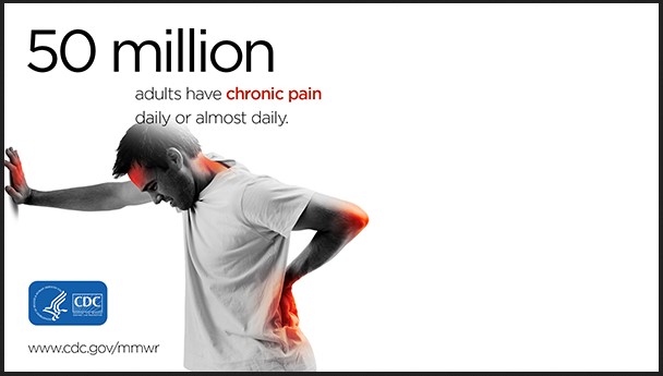
Source: cdc.gov

Chronic pain is a debilitating condition that can significantly impact a person’s quality of life. It can be caused by a variety of conditions, including arthritis, fibromyalgia, and nerve damage. Fortunately, there are a number of strategies available to help manage chronic pain and improve quality of life. This article will discuss some of the most effective strategies for managing chronic pain, including lifestyle changes, medications, physical therapy, and alternative therapies. By implementing these strategies, individuals can take control of their chronic pain and improve their quality of life.

Exploring the Benefits of Mindfulness Meditation for Chronic Pain Management

Mindfulness meditation is an increasingly popular form of meditation that has been found to be beneficial for managing chronic pain. This type of meditation involves focusing on the present moment and being aware of one’s thoughts, feelings, and bodily sensations without judgment. Research has shown that mindfulness meditation can help reduce pain intensity, improve quality of life, and reduce stress and anxiety associated with chronic pain.

The first benefit of mindfulness meditation for chronic pain management is that it can help reduce pain intensity. Studies have found that mindfulness meditation can help reduce the intensity of chronic pain by decreasing the activity of the brain’s pain-processing centers. This can help reduce the amount of pain experienced and make it more manageable.

Another benefit of mindfulness meditation for chronic pain management is that it can improve quality of life. Mindfulness meditation can help reduce stress and anxiety associated with chronic pain, which can lead to improved mood and overall quality of life. Additionally, mindfulness meditation can help reduce the amount of time spent ruminating on pain-related thoughts, which can help reduce the amount of time spent feeling overwhelmed and discouraged.

Finally, mindfulness meditation can help reduce stress and anxiety associated with chronic pain. Studies have found that mindfulness meditation can help reduce stress and anxiety by decreasing activity in the brain’s fear-processing centers. This can help reduce the amount of stress and anxiety experienced and make it easier to cope with chronic pain.

Overall, mindfulness meditation can be a beneficial tool for managing chronic pain. It can help reduce pain intensity, improve quality of life, and reduce stress and anxiety associated with chronic pain. If you are living with chronic pain, it may be worth exploring mindfulness meditation as a potential tool for managing your pain.

How to Create a Support System for Chronic Pain Management: Tips for Finding the Right People and Resources

Living with chronic pain can be a difficult and isolating experience. It can be hard to find the right people and resources to help you manage your pain and create a supportive environment. Here are some tips for finding the right people and resources to create a support system for chronic pain management.

1. Reach out to your healthcare provider. Your healthcare provider is the best source of information and support when it comes to managing your chronic pain. They can provide you with information about medications, treatments, and lifestyle changes that can help you manage your pain. They can also refer you to other healthcare professionals, such as physical therapists, psychologists, and pain specialists, who can provide additional support.

2. Connect with other people living with chronic pain. Connecting with other people who understand what you’re going through can be a great source of support. There are many online support groups and forums dedicated to chronic pain management. You can also look for local support groups in your area.

3. Utilize online resources. There are many online resources available to help you manage your chronic pain. These include websites, blogs, and podcasts that provide information about pain management, as well as online communities where you can connect with other people living with chronic pain.

4. Seek out professional help. If you’re struggling to manage your chronic pain, it may be helpful to seek out professional help. A psychologist or therapist can provide you with coping strategies and support to help you manage your pain.

5. Take care of yourself. Taking care of yourself is essential for managing chronic pain. Make sure to get enough rest, eat a healthy diet, and exercise regularly. You should also make time for activities that you enjoy, such as reading, listening to music, or spending time with friends and family.

Creating a support system for chronic pain management can be a difficult process, but it is possible. By reaching out to your healthcare provider, connecting with other people living with chronic pain, utilizing online resources, seeking out professional help, and taking care of yourself, you can create a supportive environment that will help you manage your chronic pain.

Conclusion

Chronic pain management is a complex and challenging process that requires a comprehensive approach. It is important to recognize that there is no one-size-fits-all solution to managing chronic pain, and that different strategies may be needed for different individuals. However, by utilizing a combination of lifestyle modifications, medications, physical and psychological therapies, and other treatments, individuals can improve their quality of life and manage their chronic pain more effectively.